Home

Unregelmäßigkeiten Norden Bleistift magnetic twisting cytometry Klang Üppig Schleifen

Interfacing 3D magnetic twisting cytometry with confocal fluorescence  microscopy to image force responses in living cells | Nature Protocols
Interfacing 3D magnetic twisting cytometry with confocal fluorescence microscopy to image force responses in living cells | Nature Protocols

Development and Calibration of an Optical Magnetic Twisting Cytometry for  Studying Nano-Dynamics of Living Cells | Scientific.Net
Development and Calibration of an Optical Magnetic Twisting Cytometry for Studying Nano-Dynamics of Living Cells | Scientific.Net

Interfacing 3D magnetic twisting cytometry with confocal fluorescence  microscopy to image force responses in living cells. - Abstract - Europe PMC
Interfacing 3D magnetic twisting cytometry with confocal fluorescence microscopy to image force responses in living cells. - Abstract - Europe PMC

An overview of the mechanical models for living cells. | Download  Scientific Diagram
An overview of the mechanical models for living cells. | Download Scientific Diagram

Using magnets and magnetic beads to dissect signaling pathways activated by  mechanical tension applied to cells. - Abstract - Europe PMC
Using magnets and magnetic beads to dissect signaling pathways activated by mechanical tension applied to cells. - Abstract - Europe PMC

Schematic of micropipette manipulation, magnetic twisting cytometry and...  | Download Scientific Diagram
Schematic of micropipette manipulation, magnetic twisting cytometry and... | Download Scientific Diagram

Development and Calibration of an Optical Magnetic Twisting Cytometry for  Studying Nano-Dynamics of Living Cells | Scientific.Net
Development and Calibration of an Optical Magnetic Twisting Cytometry for Studying Nano-Dynamics of Living Cells | Scientific.Net

Interfacing 3D magnetic twisting cytometry with confocal fluorescence  microscopy to image force responses in living cells | Nature Protocols
Interfacing 3D magnetic twisting cytometry with confocal fluorescence microscopy to image force responses in living cells | Nature Protocols

Stress fiber anisotropy contributes to force-mode dependent chromatin  stretching and gene upregulation in living cells | Nature Communications
Stress fiber anisotropy contributes to force-mode dependent chromatin stretching and gene upregulation in living cells | Nature Communications

In situ determination of exerted forces in magnetic pulling cytometry: AIP  Advances: Vol 9, No 3
In situ determination of exerted forces in magnetic pulling cytometry: AIP Advances: Vol 9, No 3

Application of Fluorescence Resonance Energy Transfer and Magnetic Twisting  Cytometry to Quantify Mechanochemical Signaling Activities in a Living Cell
Application of Fluorescence Resonance Energy Transfer and Magnetic Twisting Cytometry to Quantify Mechanochemical Signaling Activities in a Living Cell
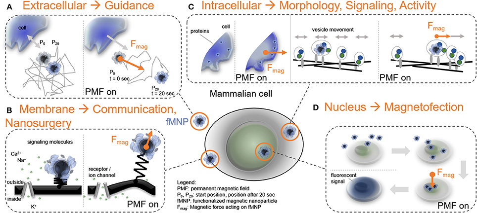
Frontiers | Force-Mediating Magnetic Nanoparticles to Engineer Neuronal  Cell Function | Neuroscience
Frontiers | Force-Mediating Magnetic Nanoparticles to Engineer Neuronal Cell Function | Neuroscience

PDF] Measurement of cell microrheology by magnetic twisting cytometry with  frequency domain demodulation. | Semantic Scholar
PDF] Measurement of cell microrheology by magnetic twisting cytometry with frequency domain demodulation. | Semantic Scholar

Interfacing 3D magnetic twisting cytometry with confocal fluorescence  microscopy to image force responses in living cells | Nature Protocols
Interfacing 3D magnetic twisting cytometry with confocal fluorescence microscopy to image force responses in living cells | Nature Protocols

Traditional experimental approaches (A) Atomic Force Microscopy (B)... |  Download Scientific Diagram
Traditional experimental approaches (A) Atomic Force Microscopy (B)... | Download Scientific Diagram

Interfacing 3D magnetic twisting cytometry with confocal fluorescence  microscopy to image force responses in living cells | Nature Protocols
Interfacing 3D magnetic twisting cytometry with confocal fluorescence microscopy to image force responses in living cells | Nature Protocols

Forschungszentrum Jülich - Expertise - Magnetic Tweezers and Magnetic  Twisting Cytometry
Forschungszentrum Jülich - Expertise - Magnetic Tweezers and Magnetic Twisting Cytometry

Micromechanical force promotes aortic valvular calcification - The Journal  of Thoracic and Cardiovascular Surgery
Micromechanical force promotes aortic valvular calcification - The Journal of Thoracic and Cardiovascular Surgery

Schematic of micropipette manipulation, magnetic twisting cytometry and...  | Download Scientific Diagram
Schematic of micropipette manipulation, magnetic twisting cytometry and... | Download Scientific Diagram

Bead-based measurements a–d, Magnetic twisting cytometry. a, Schematic... |  Download Scientific Diagram
Bead-based measurements a–d, Magnetic twisting cytometry. a, Schematic... | Download Scientific Diagram

Major techniques for cancer cell mechanics study. (a) Atomic force... |  Download Scientific Diagram
Major techniques for cancer cell mechanics study. (a) Atomic force... | Download Scientific Diagram

A: microscope stage with twisting coils and magnetization coils. B: a... |  Download Scientific Diagram
A: microscope stage with twisting coils and magnetization coils. B: a... | Download Scientific Diagram

Schematic of measurement techniques that assess meachnical properties... |  Download Scientific Diagram
Schematic of measurement techniques that assess meachnical properties... | Download Scientific Diagram

Forschungszentrum Jülich - Expertise - Magnetic Tweezers and Magnetic  Twisting Cytometry
Forschungszentrum Jülich - Expertise - Magnetic Tweezers and Magnetic Twisting Cytometry

Cell deformation and bead movement imposed by the magnetic twisting... |  Download Scientific Diagram
Cell deformation and bead movement imposed by the magnetic twisting... | Download Scientific Diagram

Forschungszentrum Jülich - Magnetic particle actuation